


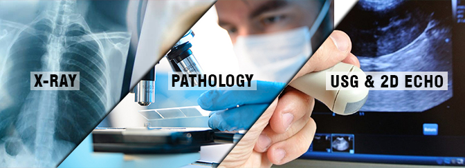
Indoor X-Ray, Pathology, USG & 2D Echo

X-Ray:
Bone X-ray uses a very small dose of ionizing radiation to produce pictures of any bone in the body. It is commonly used to diagnose fractured bones or joint dislocation. Bone x-rays are the fastest and easiest way for your doctor to view and assess bone fractures, injuries, and joint abnormalities.
An x-ray is a noninvasive medical test that helps physicians diagnose and treat medical conditions. Imaging with x-rays involves exposing a part of the body to a small dose of ionizing radiation to produce pictures of the inside of the body.
Benefits:
Bone x-rays are the fastest and easiest way for a physician to view and assess bone injuries, including fractures, and joint abnormalities, such as arthritis.
X-ray equipment is relatively inexpensive and widely available in emergency rooms, physician offices, ambulatory care centers, nursing homes, and other locations, making it convenient for both patient and physicians.
Risks:
There is always a slight chance of cancer from excessive exposure to radiation. However, the benefit of an accurate diagnosis far outweighs the risk.
The effective radiation dose for this procedure varies.
Pathology:
Pathology is a medical specialty that determines the cause and nature of diseases by examining and testing body tissues (from biopsies and pap smears) and bodily fluids (from samples including blood and urine). The results from these pathology tests help doctors diagnose and treat patients correctly. From then on we rely on blood tests, biopsies and a multitude of other pathology tests to prevent, diagnose and treat infections, allergies chronic diseases, cancers, and countless other medical conditions.
Pathologists usually work in tandem with surgeons and play a critical role in the identification of the tissue removed during the process of surgery ‑ particularly in cases when there is a tumor suspected.
2D Echo:
2D Echocardiography or 2D Echo of the heart is a test in which ultrasound technology is used to take pictures of the heart. It displays a cross-sectional ´slice` of the beating the heart, showing chambers, valves and the major blood vessels of the heart. ´Doppler` is a special element of this ultrasound exam that assesses the flow of blood in the heart. It is a preliminary test to evaluate the function of the heart and ability to throw the blood towards different body organ (EF).